Tutorial: Landmarking the left atrium
This tutorial explains how to use the landmark_atrium function to extract and separate the left atrium and left atrial appendage from 3D models of the heart. The functionality is split into two main tasks: extracting the left atrium and appendage, and separating the left atrium and appendage.
The following Python script is used to perform this operation. The script is part of the morphman package, and it utilizes various VTK-based functions for manipulating and processing heart models. We also show how to call this script from the command line using morphman-landmark.
The script accepts the following input arguments:
input_path: Path to the folder containing the model files (required).
resampling_step: The length for centerline resampling (required).
The output of the script is two separate .vtp files: one for the left atrium (LA) and one for the left atrial appendage (LAA).
The extract_left_atrium_and_appendage function detects and isolates the left atrial appendage (LAA) from the left atrium lumen based on the cross-sectional area along centerlines.
Code Reference:
def extract_left_atrium_and_appendage(input_path, resampling_step):
    """
    Detect the left atrial appendage and isolate it from the atrium lumen.

    Args:
        input_path (str): Path to folder with model files.
        resampling_step (float): Resampling length for centerline resampling.
    """
    # Load model file
    surface = read_polydata(input_path)

    # Compute centers and process centerlines
    inlet, outlets = compute_centers(surface)
    la_centerlines, _, _ = compute_centerlines(inlet, outlets, resampling=resampling_step)

    # Clip Pulmonary Veins (PVs)
    surface = clip_pvs()

    # Clip Mitral valve (MV)
    surface = clip_mv()

    # Finalize and save the clipped surface
    write_polydata(surface, input_path.replace(".vtp", "_la_and_laa.vtp"))
The separate_left_atrium_and_appendage function separates the left atrium from the appendage by clipping the regions based on a cross-sectional area along the centerlines.
Code Reference:
def separate_left_atrium_and_appendage(input_path, resampling_step):
    """
    Separate the left atrium from the appendage.

    Args:
        input_path (str): Path to folder with model files.
        resampling_step (float): Resampling length for centerline resampling.
    """
    # Load model file
    surface = read_polydata(input_path)

    # Compute centers and process centerlines
    inlet, outlets = compute_centers(surface)
    laa_centerline, _, _ = compute_centerlines(inlet, provide_point(surface), resampling=resampling_step)

    # Clip and extract the left atrium and appendage
    surface, laa_surface = clip_and_separate(surface, laa_centerline)

    # Save the separated surfaces
    write_polydata(laa_surface, input_path.replace(".vtp", "_laa.vtp"))
    write_polydata(surface, input_path.replace(".vtp", "_la.vtp"))
To run the script, you need to pass the input path of the model files and the desired resampling step size for centerline extraction.
Example:
python landmark_atrium.py --input-path /models/model.vtp
or
morphman-landmark --input-path /models/model.vtp
This will output two files in the same folder:
model_la.vtp: Contains the left atrium.
model_laa.vtp: Contains the left atrial appendage.
Below is an illustration of an atrium model having undergone landmarking.
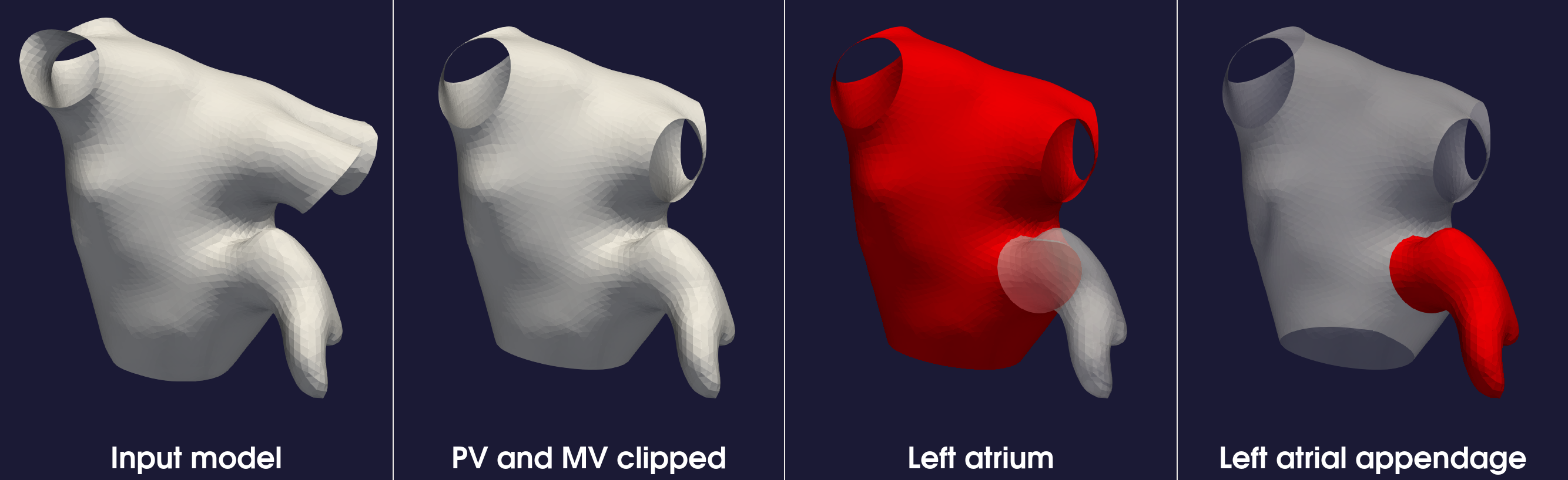
Figure 1: Steps of landmarking the left atrium and the left atrial appendage.
For more details on the algorithm used in this script, refer to the original paper by Tobon-Gomez et al. 1
For additional information, beyond this tutorial, on the script and input parameters, please run morphman-landmark --help or consult the API documentation.
- 1
Tobon-Gomez, Catalina, et al. “Benchmark for algorithms segmenting the left atrium from 3D CT and MRI datasets.” IEEE transactions on medical imaging 34.7 (2015): 1460-1473.